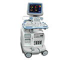
GE Vivid 7
Product Information:
Manufacturer: GE
Description:
GE Vivid 7, a premier cardiovascular ultrasound system from GE Healthcare, expands on the strength of a powerful imaging platform to offer new, innovative technology of dimensional proportions.
The easy-to use Vivid 7 significantly increases clinical productivity with time-saving features and a software-based imaging platform that fully integrates the ultrasound data throughout the entire imaging chain - from acquisition and reconstruction to analysis and reporting - for faster, more complete echo exams.
The new GE Vivid 7 Ultra-Definition Clarity Control feature provides you with multiple 2D settings that reduce noise, while keeping edges smooth. It enables the personalization of images for 4D, cardiac and vascular imaging.
Performance:
- Full integration eliminates the need to perform two separate exams, i.e., first a standard 2D exam and then a separate live 3D exam, before analysis on a separate workstation. With the Vivid 7 Dimension, the single Multi-dimensional and 4D study can be analyzed on the system or the EchoPAC Dimension workstation, dramatically streamlining both acquisition and analysis of all echo exams.
- Online 4D renderings, performed in real-time during scanning, streamline exams and save time.
- DICOM connectivity with embedded raw data storage permits post-exam quantitative analyses on the Vivid 7 Dimension system or the EchoPAC Dimension workstation; whichever best fits your echo lab workflow.
- Seamless measurement -integration allows you to efficiently calculate ejection fraction and volumes from Tri-plane images gathered from the same heartbeat.
- Blood Flow Imaging (BFI) - new vascular imaging mode gives clinicians a better understanding and delineation of directional blood flow in vessels.
- Bull's Eye Report Formats and TSI Surface Mapping - communicate cardiac dyssynchrony in a visual display that should be more familiar to EP physicians.
- 4D Tissue Synchronization Imaging (TSI) - propels Tissue Velocity Imaging to the next level by taking three simultaneous planes - from a single heartbeat at high frame rates - to create a flexible, dynamic 4D model with quantitative measurements to better communicate cardiac dyssynchrony.
- Real-time 4D Imaging - provides more cardiac information to help clinicians better communicate the heart's structure and function.
- Stress-free stress echo with Bi-plane and Tri-plane imaging enables complete acquisition of all stress echo views from just two imaging windows.
- Real-Time Full Volume - constructs a complete 4D volume online during scanning, enabling you to see the information you are acquiring in real-time, for more complete and artifact-free depictions of the entire heart.
Capabilities:
Cardiac, Vascular, General Radiology
Available Transducers:
C358, C721, Curved Abdominal, 3S, 7S, 5S, 10S Sector Adult Heart and peds Heart, 10L, 7L, Linear Vascular, 6T TEE probe
For additional informaton about this product, please call 855-951-8787 or fill out our Contact Form Today!!!